Removing liver tumors safely, noninvasively and efficiently
Many liver tumors have long been difficult or impossible to remove. Since 2015, however, it has been possible to treat these tumors by combining noninvasive surgical techniques, radiological imaging and a navigation system. For the first time, a new study by University of Bern and Inselspital, Berne University Hospital has impressively demonstrated the success of this technique.
Every year, approximately 1,250 people in Switzerland develop liver cell cancer (hepatocellular carcinoma). Among other things, this type of cancer forms small tumors that are difficult to detect and access and could therefore not be removed using conventional surgical techniques. For some years now, surgeons and interventional radiologists have had a microwave ablation method at their disposal that guides a treatment probe from the abdominal wall directly to the tumor. Without reliable planning and instrument navigation, however, this method requires excessive care because it can injure major blood vessels or even the lungs. Furthermore, the complete removal of tumors cannot be sufficiently ensured.
Innovative procedure from Bern
In 2013, the Department of Visceral Surgery and Medicine and the Department of Diagnostic, Interventional and Pediatric Radiology at Inselspital together with the ARTORG Center for Biomedical Engineering Research at the University of Bern developed a navigation system for the treatment of tumors. Thanks to the integration of magnetic resonance imaging and computed tomography, the new navigation system makes it possible to plan the access path to the tumor and guide the probe precisely to the target area by means of real-time navigation. Because the liver is constantly moving due to breathing, real-time measurement is crucial. The technology developed to market maturity by CAScination in Bern has now been introduced worldwide.
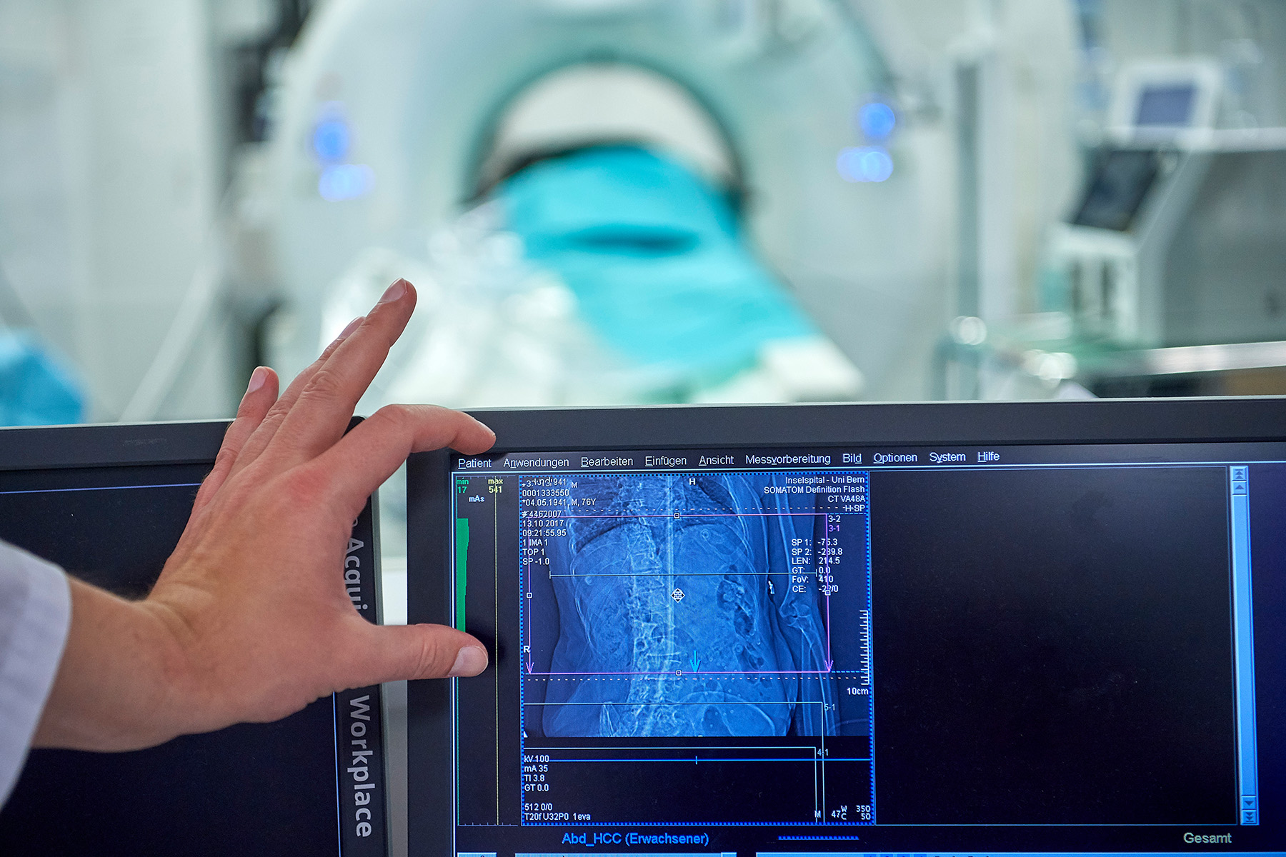
Noninvasive computer-assisted navigation to the tumor
Image-guided ablation (tumor removal) is today performed at Inselspital two to three times a week to treat patients with liver cancer as well as metastases of colon cancer and other types of cancer. Prior to the procedure, patients are safely embedded in a vacuum mattress to fix their position exactly. The system visualises the respective location of the tumors precisely in the organ before insertion and after placement of the probe, as well as for monitoring success. Navigation points on the abdominal wall and a precise target system for the treatment probe enable computer-assisted, exact implementation of the planned treatment path, as well as microwave ablation of the tumor. After the operation, the treatment team immediately sees whether the tumor tissue has been completely ablated and can make improvements if necessary.
Safe, tissue-conserving and efficient
Das Forschungsteam hat nun in einer Studie am Inselspital ausgewertet, wie sicher, therapeutisch wirksam und wirksam die Methode ist. Dazu untersuchte es 174 Ablationen von Leberzellkrebs mit bildgeführter Navigation zwischen 2015 und 2017. Die Genauigkeit der Platzierung der Sonde und der Grad der TumorentfernuAs the first user and co-developer, Inselspital has now evaluated in a retrospective analysis the method’s safety, therapeutic and procedural efficiency. To this end, the study team analyzed 174 ablations in cases of liver cell cancer between 2015 and 2017, which were carried out with the support of image-guided navigation. Needle placement and ablation coverage immediately after the procedure were checked each time in the navigation system. The results of this study were published in Liver International in October 2019. Lead author PD Dr. med. Anja Lachenmayer summarizes: “Overall, our analysis showed that we were able to efficiently remove 96.3 percent of the tumors. On average, the probe deviated only 3.2 millimeters from the ideal treatment site. The risk of complications was very low at 5.9 percent (0.9% of which were highgrade complications).” Due to the minimally invasive procedure, patients could normally leave the hospital after only two days. The head of the study emphasizes: “We are able to prove that imageguided microwave ablation is a safe, tissue-conserving and efficient treatment for removing liver tumors. With the new system, we can even detect tumors not visible with conventional imaging and also treat areas that have remained inaccessible until now.” The latter is crucial for many patients because in more than half of liver cell cancer cases, tumors are untreatable because of difficult anatomic locations and could not be treated without navigation support.
Focus on Bernese technology at International ECALSS Meeting 2019With nearly 600 liver tumors treated (in 391 interventions, 590 tumors removed), Inselspital has some of the world’s greatest experience with the ablation navigation system that was developed in Bern. Visceral surgeon Anja Lachenmayer and interventional radiologist Martin Maurer presented their experiences with the navigation system to international specialists in surgery, radiology, biomedical engineering and in the industry at the annual meeting of the European Computer Assisted Liver Surgery Society ECALSS (October 17-19, www.ecalss.org). |
Publication details:Anja Lachenmayer et al.: Stereotactic image‐guided microwave ablation of hepatocellular carcinoma using a computer‐assisted navigation system. Liver International, October 2019, https://doi.org/10.1111/liv.14187 |
Source: Inselgruppe
2019/10/31