Software detects at-risk tissue in record time following a stroke
The FASTER software developed in Bern can detect within minutes the areas of the brain that will be left with long-term damage following a stroke. The previous version – BraTumIA for tumour segmentation – has been in use around the world since 2014.
In October of last year, a fully automatic computer program for the detection of brain tumours, which was developed in Bern, caused something of a stir on the international stage. The BraTumIA software only needs 10 minutes to analyse the tissue structures within a malignant tumour in very great detail. The self-learning system was developed and validated by biomedical engineers at the University of Bern's Institute for Surgical Technology and Biomechanics (ISTB) in collaboration with neuroradiology consultants at the Inselspital. The software has been used by more than 200 users in over 40 countries since its release (May 2014).
From brain tumour to stroke
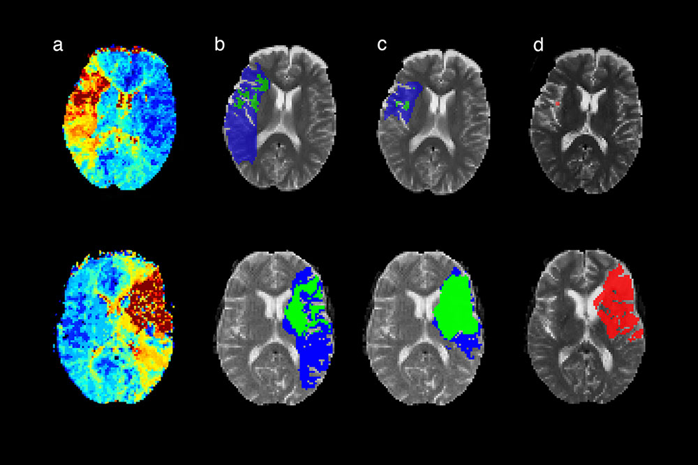
Drawing on the analysis mechanisms and experiences associated with BraTumIA, the team has now developed a new type of software that identifies areas of the brain that might be at risk following a stroke. The really clever thing is how it only takes the computer 6 minutes both to detect any tissue affected by a direct loss of perfusion and to predict which areas of the brain will probably be left damaged after an intervention. This information enables doctors to identify more precisely which tissue has a chance of complete recovery and then free this in a targeted manner using a catheter. The system bases its risk assessment on pre-learned realistic scenarios.
First place for independent imaging
On 5 October, the new software known as FASTER achieved first place (for stroke-related imaging processes) in the international ISLES challenge held during the MICCAI international biomedical conference. The software was developed by Dr Richard McKinley, a mathematician and academic based at the Support Center for Advanced Neuroimaging (SCAN) within the Neuroradiology Department at the Inselspital. 'The close collaboration between the neuroradiologists at SCAN and the engineers at ISTB was crucial to winning this competition,' explains McKinley. 'Our approach combines precise algorithms, modern imaging and clinical expertise.'
From Bench to Bedside
The system operates independently, is constantly learning at the same time, and can be 'trained' by experienced clinicians to characterise strokes at lightning speed using MRI images. This directly improves treatment for patients – one of the stated aims of the Swiss Institute for Translational and Entrepreneurial Medicine (sitem-insel AG) in Bern. The research group is already working on a new type of software for the analysis of inflamed brain tissue in multiple sclerosis patients.
2015/10/28